Pots and Pieces
The Anatomy Museum of the Otago Medical School
and how it came to be
The lively environment of the modern day Anatomy Museum would probably shock some of the people who were responsible for its existence. They would see students from a variety of nonmedical courses, in addition to medical students, freely able to handle the specimens and models and using the room as a classroom and resource centre. They would also see artists at work and school children on class trips. The equal proportion of men and women using the museum would probably also surprise.
The Anatomy Museum showing the classical statues bought by Professor Scott in the 1880s
|
Each year about 800 students from Otago University and Polytechnic have regular classes in the Museum and another 400 use its resources in other locations within the Department of Anatomy and Structural Biology. The Museum is not open to the public but about 40 school and special interest groups make escorted visits if they can find a gap in the timetable. The Museum’s resources include about 2,000 catalogued specimens and models illustrating mostly normal human anatomy, a similar number of radiographs, a variety of permanent and semi-permanent displays, videos and audiotape programmes, and various other teaching materials.
Amongst all of this modernity and activity the Anatomy Museum still has many reminders of its past. The room is filled with fine woodwork and natural light from the Edwardian-style skylights. Replicas of classical statues stand atop a glass case containing rows of fine plaster models, while around the room are other unmistakable examples of model-making from the late 19th and early 20th centuries. Old and new combine with the ever-present fascination for human anatomy to produce a truly unique environment.
The Museum’s early history was bound up with the establishment of the Medical School at the University of Otago and owed a great deal to the Professors of Anatomy who almost singlehandedly brought it into existence. The University itself was opened in 1871, only 23 years after the first Scottish colonists arrived in Otago and ten years after the Otago goldrush. Millen Coughtrey, aged 26, was appointed as the first Professor of Anatomy and Physiology in 1874, and classes for the first four medical students started the following year. The whole of the University at the time was in a building in the Exchange, in central Dunedin, which later became the Stock Exchange and was eventually pulled down to make room for John Wickliffe House. There are no records of an Anatomy Museum from that time but specimens from dissections would have been preserved, and one can imagine enthusiastic local doctors providing curiosities for the fledgling Medical School.
The first few years were rather shaky. Coughtrey gave public lectures around Otago to popularise the need for a Medical School, but insufficient planning resulted in a failure to gain essential recognition from the “Home” Universities. When Coughtrey resigned in 1876 because he was not allowed to treat patients and so make a reasonable living, the whole idea of a Medical School was almost dropped. However, James Macandrew, the Superintendent of Otago, was convinced of its importance to the new colony and eventually won over the University Council to try it again.
John Halliday Scott, another 26-year-old, was appointed Professor of Anatomy and Physiology in 1877 and so began a long period of growth for the Medical School that lasted for thirty-seven years. Dr Scott had a strong Scottish character and had a “genius for order and method”. Under his leadership and hard work the Medical School rapidly made progress. Scott was a watercolour artist and former students commented on his beautiful and clear blackboard drawings, which they found painful to rub out at the end of the day. He painted wall charts, still used to this day from material in the dissecting room and took photographs of dissections when photography was in its infancy.
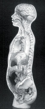
Fine plaster cast of the internal organs acquired during the 1880s
|
The Medical School was moved to the freshly built blue stone buildings by the Leith stream in 1879, into rooms now occupied by the Geology Department. In 1881, Professor Scott obtained a grant of 20 pounds specifically for the Anatomy Museum. In 1892 a large number of models were presented to the Medical School by Dr Maunsell, a retired lecturer in Surgery. A large group of models in the Museum date from this period. These include the plaster torso and head models from the Leipzig firm of Steger as well as replicas of classical statues by D. Brucciani and Co. of London. The Anatomy Museum statues include “Venus de Milo”, whose original stands in the Louvre; “Lorenzo de Medici” by Michaelangelo; the “Borghese Gladiator” whose detailed venous and muscle markings suggest that dissection was practised in Greece as early as 100 BC; and a dissected human body that was possibly made from drawings done by Vesalius in his book De Humani Corporis Fabrica published in 1543. Several of the wax models of developing embryos may also have been obtained toward the end of the 19th century.
During the whole time that Scott was head of the Anatomy and Physiology Department and later Dean of the Medical School, he had only one assistant, the remarkably versatile but dour Alfred Jefferson. Jefferson was the dissecting room porter or “corpses” friend. He also prepared specimens and models, and carried out odd jobs. In the memoirs of a former medical student he was described as “capable and obliging but appeared rather morose, as if under a perpetual grievance”. At least 130 models and wet preparations in the present Museum would have been produced as a collaboration between Scott, the anatomist, and Jefferson, the technician. In those times all medical specimens were stored together but when the Pathology Department was established Anatomy and Pathology developed separate museums.
In 1914 Scott died and not long after Jefferson resigned, bringing to a close a long period of hard work and development for the Medical School and its Museum. The following phase was dominated by Professor William Percy Gowland who had trained as a doctor in London. He brought a different style to the Otago Medical School, and with it implications for the Anatomy Museum. “A Lancashire Lad”, with a very loud voice, he was devoted to teaching elementary anatomy, and he also inspired much research in the field of anatomy. He employed a series of medical graduates, who served as demonstrators in classes and carried out research projects. Many of these projects resulted in the preparation of wet specimens or models for the Museum. Gowland also increased the staff of the Department significantly by employing technical officers, secretaries and an artist, in addition to the anatomy steward who cared for the bodies in the dissecting room.

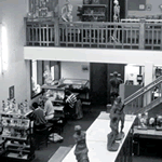
The Anatomy Museum as it was in 1927
|
The decision to build the Lindo Ferguson building was made in 1919 because of overcrowding by ex-soldiers enrolling in medicine after the First World War. Eight years later, in 1927, the Lindo Ferguson building was finally completed. Several cost-cutting measures were made to keep within budget, such as an absence of fire escapes, a dangerous situation not remedied until 1959. Transferring the Museum specimens, lab equipment, furniture, and cadavers from the old Medical School was a big job. The man who normally transported the cadavers for the Department with his one-horse carrier van, had to be convinced with quite a lot of whisky, at the Department’s expense, to help out on the day.
In the purpose-built Anatomy Museum, displays were kept in large glass cabinets and were not accessible to students. The mezzanine floor housed the big Scott collection of Mäori and Moriori bones and was closed to students. Specimens from the museum were used in dissecting room classes but no regular classes were held in the Museum. Former students remember the Museum as rather forbidding, in part because its most significant use by them was during oral examinations. It was a Museum in the old-fashioned sense of the word, quiet, still and impressive, and it stayed that way right up until the 1970s. Wax models such as a series of dissected heads and necks of bewhiskered Edwardian gentleman, were made by Thomas H. Kelsey, a highly skilled modeller and artist. Most of his work was produced between 1927 and 1929, when improvements were carried out in the Museum. Better shelving, lighting and new museum jars were required to fill the teaching demands of several newly established clinical departments. Mr Kelsey worked under the supervision of Dr Durward and used special techniques, which he guarded well.
Gowland would have reached the statutory retirement age of 65 in 1944, but he retired in 1943 when the Government decreed that the medical intake would be increased from 100 to 120 students per year. Gowland had previously reported overcrowding in his department, and when his protest at the latest increase was not supported by the University Council, he resigned.
William Edgar Adams did his MSc at Otago and then went on to do medicine as a student of Professor Gowland. He worked as a senior demonstrator at Otago and was later lecturer at Leeds University. He returned to New Zealand to take over the Chair of Anatomy when Gowland resigned. Adams was Head of Department from 1944 until 1969 when he was appointed Dean. His research field was neurology and this led to a series of plaster models of brain dissections in the Museum. He was a traditionalist and maintained the Department and its Museum much as it was under Gowland. During this period, though, the subject of Anatomy began to be taught to students other than medical students, starting with those from physical education and dentistry. Although women had been admitted to the University from its inception, women medical graduates, the first of whom was Emily Siedeberg in 1896, were rare in the early years. It was not until the 1950s that significant numbers, about 10 per cent, regularly appeared in Anatomy classes.
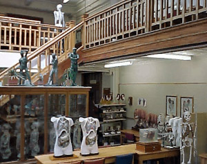
The modern day Anatomy Museum
|
William (Bill) Trotter was an anatomy demonstrator from 1947 to 1948 and was on the Anatomy staff from 1949. In 1969 after Adams’ appointment as Dean, Bill became Professor of Anatomy, retiring in 1983. One of his major achievements was turning the Anatomy Museum into a classroom containing several smaller tutorial rooms and also getting rid of nearly all of the locked glass museum cases, thereby making the models and specimens accessible to students. The major rebuilding started in 1972 and was not completed until 1982.
Three technicians who joined the staff during Gowland’s and Adams’ time had a profound influence on the museum. Keith Pickersgill was a technician from 1945 until he died suddenly in 1988. He helped remove specimens from their big glass cases, made perspex museum jars and models in fibreglass and plaster. Charles Unwin was anatomy steward from 1958. From 1972 to 1978 he worked exclusively in the museum. He was a skilled cabinet-maker and made many fine wooden bases, containing drawers with keys for models. Margaret Ogilvie painted keys and diagrams for the models, and repainted some of the older plaster models. Associate Professor Len Robinson, who joined the staff in 1952, took special responsibility for the Museum and worked with the three technicians on the production of models and their keys.
Models from the French firm Auzoux made their way into the Museum around the time of the renovations. Of note were the Nicholas-Augier-Roux models, which consisted of fibreglass casts of dissections. These were unusual because they were from young healthy people – criminals who had been given the death penalty. Russell Barnett has been the Museum Preparator since 1979. He has a special talent for making fibreglass models. He also uses the very modern technique of plastination, which preserves anatomical specimens without having to keep them in fluids. The process involves the gradual replacement of the water in a specimen with solvents and then silicone or epoxy plastic.
The current Head of Department is Professor Gareth Jones, who has held the post since 1983. He has very much fostered the use of the Museum as a classroom and resource centre. In the last few years the development of Anatomy as a science subject and the growth of research into cellular anatomy using electron microscope techniques has led to the Department’s name change to that of the Department of Anatomy and Structural Biology. With Physiotherapy becoming a degree course, increases in the number of physical education students, and Anatomy classes for Polytechnic courses such as Occupational Therapy, the Department has grown, and it is now one of the largest in the University.
I am not sure what the future holds. Rapid changes in the structure of the University in recent times and increasing student numbers mean that the Anatomy Museum is by no means at the end of the road. Many medical schools in Australia and England lost their old-style museums when they restructured but are now, several years on, regretting the resources that they lost. This Anatomy Museum is still in existence because it did not remain a static display of curiosities but instead adapted to the changing needs of students.
This is a modified version of an article written by Fieke Neuman, the full version of which can be found in the New Zealand Museum’s Journal (1993) 23(1):17-22.
What follows is a brief update of the life of the Anatomy Museum in the intervening years between 1993 when the above article was written, and the present day. The article’s author was Fieke Neuman, who was the Museum Curator from 1988 to 1996. Fieke resigned in 1996 in order to a pursue a career as a designer in the fashion industry. She was responsible for computerising the Museum catalogue (an important aid to staff and students), and undertook the development of the projection slide catalogue. The current Curator of the Museum is Tracy Connolly, who started work in the Department in 1996. Tracy is a graduate in Anthropology and aims to promote modern archiving standards and care of specimens within the Museum environment.
The Anatomy Museum has continued to adapt to changing times. It now caters for well-over 1250 students in disciplines as diverse as Medicine, Dentistry, Science, Medical Laboratory Science, Nursing, Physiotherapy, Physical Education, Design Studies, Biological Anthropology and Neuroscience. The number of catalogued specimens has also increased from 2000 in 1993 to 2500 in 2004. The vast majority of specimens that have been added to the collection in more recent times are plastinated. The Department is fortunate to have retained the services of Russell Barnett who has honed his plastinating skills to a fine art. Pride of place in the Museum’s collection must go to “GI Joe”, a vertical, life-size, plastinated dissection of the gastrointestinal system from start to finish. A series of plastinated thin sections through the human brain also represent something that would have been almost unimaginable 100 years ago.
In 2002, the Museum was renamed the W.D. Trotter Anatomy Museum in honour of Professor Trotter who passed away in 2001. Professor Trotter was instrumental in enlarging the museum and removing models from locked cabinets, thus providing students with a valuable hands-on approach to learning.
Professor Gareth Jones stepped down as Head of Department in 2003 and the position was filled by Professor Helen Nicholson, from the University of Bristol. Professor Nicholson’s expertise and interest in a wide range of disciplines, including medical teaching and clinical anatomy will ensure that the Anatomy Museum continues to go from strength to strength. The current prediction is that its treasures will form a vital facet of students’ education for many years to come.
Fieke Neuman, Gareth Jones and Kerry Galvin

|