This page describe the techniques our scanning electron microscopes can provide. Visit Transmission electron microscopy techniques for TEM techniques.
Array Tomography
Resin-embedded tissue is serial sectioned onto a glass microscope slide. The sections are then stained with a fluorescent dye or tag and imaged with the confocal microscope. The sections can then be eluted and restained with another fluorescent tag or dye. After the desired labels have been imaged the sections are stained with a heavy metal and then imaged using backscatter electron microscopy. A three-dimensional model of the antigens, fluorescent proteins and ultrastructure in individual tissue specimens can then be constructed.
Backscattered Electron Imaging (BSE)
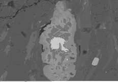 This technique is used to detect contrast between areas with different elemental compositions, as well as, surface topography. Higher atomic number material appears brighter than low atomic number material in a backscattered electron image. BSE images are very helpful for obtaining high-resolution compositional maps of a sample and for quickly distinguishing different phases. They are often used in conjunction with spot probe analyses by energy-dispersive x-ray spectroscopy.
Backscattered electron imaging can be performed on our Zeiss Sigma VP FEG SEM, JEOL FE-SEM6700, Cambridge 360 SEM.
Cathodoluminescence (CL)
This provides information about the trace elements (typically transition metals or rare earth elements), contained in minerals and can be used to create compositional maps. It also provides information about processes such as crystal growth, replacement, deformation, provenance and the production of mechanically-induced defects. It is most commonly used in geological studies.
Cathodoluminescence imaging is not available at present, but we hope to offer this service soon. Contact allan.mitchell@otago.ac.nz for the latest advice.
Cryo-SEM
 Samples are solidified by plunge freezing in nitrogen and can withstand the vacuum conditions required for examination in the SEM while kept at low temperatures on the cryo-stage. This allows samples to be viewed in their hydrated state and is particularly useful for liquid, semi-liquid and beam-sensitive samples. There is usually no exposure to toxic reagents and little mechanical damage to samples. Cryo-SEM is the most effective method of preventing sample water loss and retaining soluble materials, meaning there is less relocation of highly diffusable elements. Cryo-SEM can be used to study dynamic processes using a series of time resolved samples. Our JEOL FE-SEM 6700 has a cryo preparation stage and sample chamber.
Electron Backscatter Diffraction Imaging

Sheared rock by David Prior.
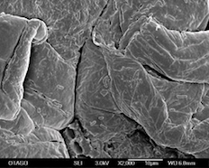
This is used to determine crystal structures and orientations of minerals, which can be used to elucidate texture or preferred orientation of any crystalline or polycrystalline material. Applications include crystal orientation mapping, defect studies, phase identification, grain boundary and morphology studies, regional heterogeneity investigations, material discrimination, microstrain mapping, and physico-chemical identification (using complementary SEM techniques).
Electron backscatter diffraction imaging can be performed on our Zeiss Sigma VP FEG SEM.
Image of sheared rock by Professor David Prior
Electron backscatter diffraction (EBSD) map of a sheared rock made mostly of grains of the mineral plagioclase. The red to green colour of each pixel shows the orientation. Yellow points are a different mineral (hornblende). Tone underlying colour shows changes in EBSD pattern quality. Blue lines are twin boundaries, where the crystal axes change by 180 degrees.
Energy-Dispersive X-ray Spectroscopy (EDS or EDX)
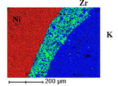 An analytical method used for determining the elemental chemical composition of a sample, and to create element composition maps over a broader area. Element maps display element distributions in textural context, particularly for showing compositional zonation. EDS can either be used for qualitative or quantitative analysis. Qualitative analysis is used to determine which elements are present in a particular location. Quantitative analysis is used to find out the relative amounts of the elements, or how much of an element is present. Line profile analysis can also be performed with EDS.
Energy-dispersive x-ray spectroscopy can be performed on our Zeiss Sigma VP FEG SEM, JEOL FE-SEM6700. Features or phases as small as a micron can be analyzed (sample dependent).
High Resolution SEM
The secondary electrons generated as the electron beam enters the specimen (SE1), are localised within a few nanomenters of the impact sites of incident high-energy electrons and can respond to local fine-scale features. The SE1, in combination with BSE (backscattered electron imaging), that retain a large fraction of the beam energy, is considered a 'high resolution' signal. High resolution SEM aims to either separate the high-resolution signals from the low-resolution signals, or use operating conditions that create similar ranges for all of the signals. We are able to perform high resolution SEM on our JEOL FE-SEM 6700 and Zeiss Sigma VP FEG SEM.
Secondary Electron Imaging
Volcanic lake sediment.
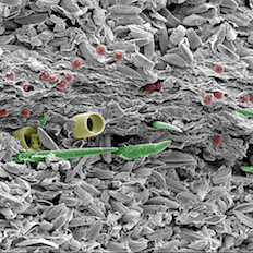
This technique provides high-resolution imaging of fine surface morphology.
Secondary electron imaging can be performed on our Zeiss Sigma VP FEG SEM, JEOL FE-SEM6700, Cambridge 360 SEM (up to 150,000 sample dependent).
Image of volcanic lake sediment by Uwe Kaulfuss
This image shows finely-layered sediment from a 23 million year old volcanic lake in Middlemarch, Otago. Light-coloured summer layers of pennate diatoms are interbedded with a winter layer of plant debris, centric diatoms (yellow), freshwater sponges (green), and algal resting spores (red). Colours in this scanning electron microscope image are artificial.
Variable Pressure SEM (VP SEM)
Conventional electron microscopes require that the electron-optic column and specimen chamber be under a high vacuum so that the electron beam can travel from the source to the sample without being scattered by residual gas atoms. This means that samples that are unable to withstand being under vacuum, or samples that would contaminate the vacuum system are unable to be imaged in conventional microscopes.
Variable pressure scanning electron microscopes are able to operate with the specimen chamber at high and low pressures. Our new Zeiss Sigma VP FEG SEM is able to image specimens at pressures from 2 to 133 Pa. This allows samples that would otherwise be unsuitable for observation to be imaged. For example porous materials such as bone, wet or damp specimens, and biological specimens that cannot be maintained in their original state if they are allowed to dehydrate. The gaseous environment allows poorly conducting and insulating samples to be imaged without the need to coat them with a conducting metal layer. The gas in the specimen chamber also creates a microenvironment which can be used for a wide variety of in situ experiments (e.g. corrosion and oxidation).
Exhibition images
See the images some of our talented staff produced:
2018 Science Festival photo exhibition