OMNI staff created some stunning and prize-winning images that were exhibited in the 2018 International Science Festival. View the images and read about how they were created.
Read more about the winners:
Science Festival: Biomedical Sciences Photo Competition impresses (Otago Bulletin)
Liz Girvan: Winner, Professional Staff category
Liz submitted three entries:
Outer space
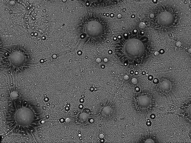
This space-like scene is from a small area inside the glass from a blown fuse. The fuse came from a power supply that ran part of the same microscope that I used to take the image. It was taken on a JEOL 6700F Scanning Electron Microscope.
The image was taken using a backscatter detector. This detector is used to show any compositional differences within the sample. The little spheres are made up of copper and lead. They have come from the metal wire in the fuse.
They show up brighter than the darker glass background as they are higher in atomic number.
This image came about as I was curious what the inside of the blown fuse would look like. I didn't expect to see something so beautiful from a fuse that had caused me so many problems with my microscope!
Trapped
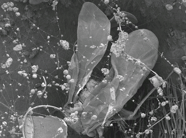
I was searching a plant sample for something completely different when I came across this spider's web. Trapped in the spider's web was this tiny insect. The sticky strands of the web have also trapped bits of dust and pollen. It was taken on a JEOL 6700F Scanning Electron Microscope.
Winter is coming (winning entry)
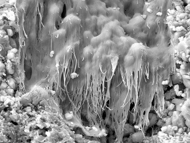
I am actually not sure what this wee structure is. It looked to me like some kind of frozen waterfall, it really stood out amongst a sea of mineral crystals. It's pretty small – about the width of a human hair.
It was inside a small piece of limestone I picked up while on a walk on the family farm. I am always picking up bits and pieces to look at under the SEM. Looking at unknown samples is a really good way of trying new techniques on the SEM. Also, you always find amazing and beautiful structures!
OMNI expertise
 Liz Girvan is a member of OMNI's Electron microscopy team. Her expertise is in Scanning Electron Microscopy (SEM) including:
- Conventional SEM sample preparation and imaging
- Field emission SEM sample preparation and imaging
- Cryo - FESEM
- Energy dispersive x-ray analysis (EDS)
Email liz.girvan@otago.ac.nz
Sharon Lequeux
Sharon submitted two entries:
Lung cancer cell undergoing mitosis
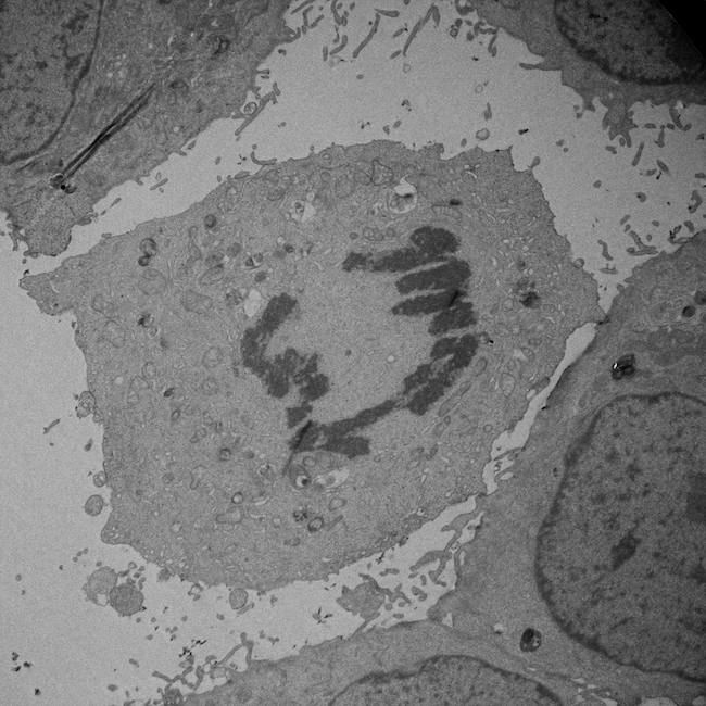
TEM micrograph taken on a JEM-2200FS Cryo-TEM at 2000x magnification. The chromosomes have condensed and the sister chromatids will be pulled towards opposite ends of the cell.
Gut organoid
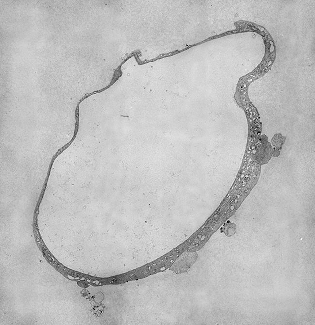
A four-day old mini-gut cultured from an intestinal stem cell. As they age these organoids grow complex structures in culture that closely parallel their in-vivo counterparts. Tissue provided by the Butt Lab (Microbiology). Micrographs were taken at 2500x on a JEM2200FS Cryo-TEM and montaged using IMOD and Photoshop.
OMNI expertise
 Sharon Lequeux's expertise in Electron Microscopy includes:
- Correlative microscopy
- Immunolabeling
- Ultramicrotomy
- Cryo-ultramicrotomy
- High pressure freezing and plunge freezing
- Conventional TEM and SEM sample preparation and imaging
Email sharon.lequeux@otago.ac.nz
Andrew McNaughton
Look head
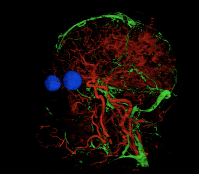
The veins and arteries were selected from a series photographs of horizontal slices taken through a human head. Rather than laboriously tracing each one, open source machine learning software (Ilastik) was taught to recognise the veins and arteries. The software then traced through all the slices to reveal the network of vessels running through the brain and parts of the face. The eyes were added in a similar manner to help show the overall context.
This is a single frame from a 3-dimensional model which can be rotated to reveal the network of vessels from any angle.
OMNI expertise
 Andrew McNaughton works with confocal and light microscopes, and the X-ray µCT. Consequently, he has developed a diverse range of interests and is developing more of the image analysis aspects of data acquisition from these instruments. He also uses 3D reconstruction of sometimes large data sets.
Confocal microscopy in particular is capable of producing some remarkable images.