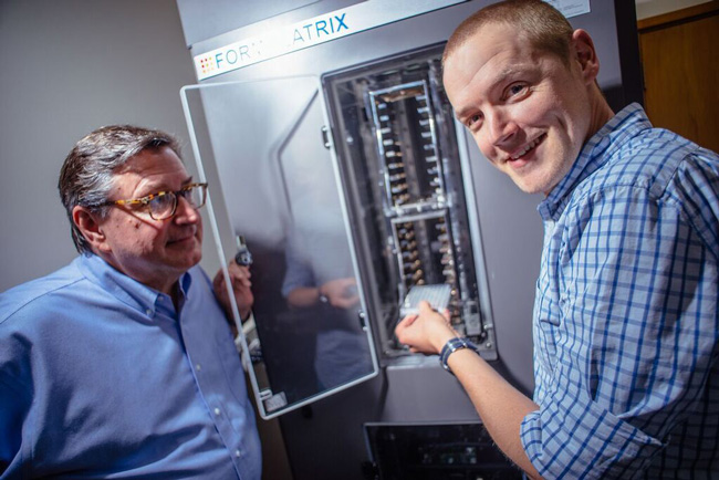
Professor Kurt Krause (left) and Dr Peter Mace of Biochemistry with the University's newly installed protein crystal imager. Photo: Alex Lovell-Smith.
A recently installed automated system for growing and imaging protein crystals is set to significantly boost structural biology research at Otago.
The new state-of-the-art unit, which is based in the Department of Biochemistry, was obtained with the aid of a Major Equipment grant from the University. It will be used by more than a dozen research teams across several departments.
Biochemistry's Dr Peter Mace and Professor Kurt Krause have overseen the facility's establishment. Dr Mace says protein crystallography is a sophisticated technique used widely in drug and vaccine design and development, which involves forming crystals from proteins that can then be x-rayed to determine the protein's 3D structure.
Protein crystallography's growth at Otago
The technique's use at Otago has expanded rapidly in the past decade and has grown from three to 14 research groups. Most of these teams are employing the method to understand complex biological systems underlying human disease.
These systems range from proteins that cause improper cell growth in cancer, inflammatory disorders, and developing new treatments for tuberculosis as well as other bacterial and fungal infections.
Professor Krause says understanding the exact biochemical properties of cellular systems in both healthy and diseased states is vital for the development of new therapeutics.
“It also allows us to understand the mechanism of action of existing therapeutic agents in order for them to be used more effectively,” he says.
Automating a time-consuming and challenging task
Dr Mace says that in a typical crystallisation experiment researchers will test a protein against around 500 or more chemicals, after which each experiment must be checked individually in a regular manner.
“This has traditionally been done by hand with a standard microscope. But now we are tackling projects on a scale that is really hard to keep track of that way,” he says.
Aside from the time and physical challenge of observing thousands of microscopic experiments, manual observation misses transient crystals and tends to falsely report positive crystals, he says.
"This isn't really surprising. We are human, each crystallisation experiment is a tiny drop about 2mm across, and 'good' crystals that do grow are normally less than 0.1mm long."
Now, with the new unit, Otago researchers are able to simultaneously keep 190 plates in a temperature controlled environment, and automatically capture images of every experiment on any fixed schedule. This capacity means users can track up to 50,000 individual crystallisation trials, and check their progress from anywhere in the world over the internet.
New imager's impact already felt
Although it was only installed a few weeks ago, it is already having an impact.
"The beauty of automated imaging is we can catch crystals that grow then disappear, or tell when a crystal has stopped growing and is ready to be harvested," Dr Mace says.
"We have already caught crystals that we would have missed otherwise, and now have new leads to follow up. I suspect students will really appreciate not having to spend all those hours looking down a microscope, leaving all the more time for planning the next experiment!"
Additional support for the imager was provided by a Lotteries Health Research grant, and contributions from the Department of Biochemistry, Department of Microbiology and Immunology, School of Pharmacy and Faculty of Dentistry, as well as the Otago School of Medical Sciences and the Division of Health Sciences.