 Monday 3 February 2014 10:06am
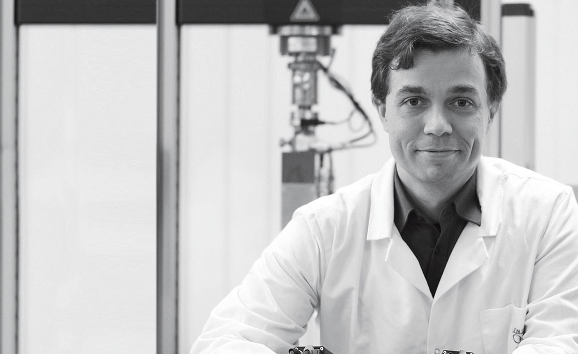
Dr Niels Hammer is a modern anatomist, using highly specialised technologies to discover new information and to help develop new surgical techniques.
Having surgeons walk across the street from the hospital to his department to discuss their surgical observations is something Dr Niels Hammer (Anatomy) values greatly about research at the University of Otago.
"When clinical colleagues come around is usually when clinical anatomists can likewise learn applied anatomy," he explains.
One recent example involved neck surgery where a surgeon located a particularly distinctive nerve – the vagus nerve – but found it wasn't exactly where the textbooks said it should be.
Hammer and his colleagues were able to turn to what he describes as a "wonderful resource" – the bodies that have been generously donated for teaching and research. By carrying out a series of dissections they found the nerve is not as constant a structure as the textbooks indicated.
"What we came across was a significant variation involving a branching pattern of the nerve. The branching of this nerve in this particular neck region had not been described at all before, so that was quite a novel finding," he says.
"This is a very applied part of research because it immediately translates into practice in the hospital across the road. This is only made possible by the gift-of-body donation."
Hammer came to Dunedin just over a year ago from Leipzig in Germany where he had worked as both a medical doctor and surgeon before focusing fully on research.
This surgical background was an excellent preparation for his clinical biomechanics research, designed to provide clinicians with very specific background information for inventing new surgical procedures, or improving existing techniques to improve patient outcomes.
"Surgery has to involve not only the topographical, spatial relationship knowledge and morphological knowledge, but also the functional knowledge that anatomy researchers can feed in as well."
It helps surgeons to not only understand where a structure is, but also what might happen if it has to be transected during an operation.
"One of the principles in surgery is to not transect what you don't need to. Sometimes – if structures are in the way – you have to."
Although light microscopy and dissection remain key ways to examine anatomical structures, Hammer is very much a modern anatomist, using specialised techniques for his research and teaching.
Computed tomography and magnetic resonance imaging (MRI) enable detailed images of tissues, providing new opportunities to answer clinicians' questions. Another approach is plastination – in which the water in biological tissues is replaced by a resin.
"If you combine these techniques of clinical imaging and plastination, you can create views into the human system that have not been generated before. The knowledge gain is even further enhanced by electron microscopy."
The biomechanical testing Hammer does also involves imaging. Here he uses stereo camera systems to help capture how anatomical structures with surgical implants or tissue-engineered scaffolds deform three dimensionally when put under a load.
The combination of blue-skies research, clinical research and teaching excites Hammer, especially when it is so closely tied to the work of hospital clinicians.
"The clinicians are very keen on interacting with Anatomy because they see it as a resource, and also as a good partnership for collaboration. That knowledge influx helps us keep the teaching content for medical and anatomy students up to date," he says.
"The proximity to the clinical side is of huge benefit and a privilege that most anatomy departments throughout the world – and I've been in many places – don't have. It is something unique about this campus."
Funding
- MedTech CoRE
- Otago School of Medical Sciences (University of Otago)
- Federal German Ministry of Economic Affairs and Energy