You are free to use the videos, photos, software and datasets below, but please refer to the relevant publication or give credit to the authors.
| Videos | Datasets and software | Photos |
Videos
Larva of the hairworm Gordius sp. (Nematomorpha: Gordiidae) extruding its three hooked rings and proboscis following excystment (filmed by Jean-François Doherty)
Download the video.
Metacercariae of the eyefluke Tylodelphys sp. (Trematoda: Diplostomidae) in the eye of a common bully, Gobiomorphus cotidianus (filmed by Isabel Blasco-Costa)
Download the video.
Explosive excystment of the metacercaria of Apatemon sp. when placed in host bile, in both normal speed and in slow motion (filmed by Bronwen Presswell)
See Blasco-Costa, Poulin and Presswell (2016) Parasitology Research 115: 271-289
Download the video.
Metacercariae of Apatemon sp. immediately following their emergence from cysts (filmed by Bronwen Presswell)
See Blasco-Costa, Poulin and Presswell (2016) Parasitology Research 115: 271-289
Download the video.
Cercariae of Curtuteria australis penetrating the foot of their bivalve host, the New Zealand cockle Austrovenus stutchburyi (filmed by Tommy Leung)
See Leung and Poulin (2011) International Journal for Parasitology 41: 449-454
Download the video.
Redia of Philophthalmus sp. feeding on a sporocyst of Maritrema novaezealandensis (filmed by Tommy Leung)
See Leung and Poulin (2011) International Journal for Parasitology 41: 1063-1068
Download the video.
Close-up of redia of Philophthalmus sp. feeding on a sporocyst of Maritrema novaezealandensis (filmed by Tommy Leung)
See Leung and Poulin (2011) International Journal for Parasitology 41: 1063-1068
Download the video.
Redia of Philophthalmus sp. feeding on a cercaria of Maritrema novaezealandensis (filmed by Tommy Leung)
See Leung and Poulin (2011) International Journal for Parasitology 41: 1063-1068
Download the video.
Datasets and software
Data on the geographic coordinates where 4943 helminth species have been discovered, from Poulin and Jorge (2019) Parasitology 146: 168-175 [Dataset]
Data on densities of 310 populations of parasitic metazoans and those of their hosts in lake ecosystems, from Lagrue and Poulin (2015) Oikos 124: 1639-1647 [Dataset]
Data on body masses and densities of free-living and parasitic metazoans from lake ecosystems, from Lagrue et al. (2015) PNAS 112: 1791-1796 [Dataset]
Data on aggregation levels of helminths in 410 samples of fish hosts, from Poulin (2013) Parasitology 140: 541-546 [Dataset]
Data on morphometrics of transmission stages from 404 trematode species, from Koehler et al. (2012) Evolutionary Ecology 26: 1497-1512 [Dataset]
Data on parasite species richness and host body size in host species from 6 taxonomic groups, from Poulin et al. (2011) Oikos 120: 740-747 [Dataset]
Data on taxonomic resolution in 950 surveys of helminth communities in 650 species of vertebrate hosts, from Poulin and Leung (2010) Parasitology 137: 1967-1973 [Dataset]
Data on species richness of cestodes in 326 species of elasmobranchs, from Randhawa and Poulin (2010) Ecography 33: 866-877 [Dataset]
Data on 106 experimental metacercarial infections, from Poulin (2010) Parasitology 137: 889-898 [Dataset]
Data on 145 experimental cercarial infections, from Poulin (2010) International Journal for Parasitology 40: 371-377 [Dataset]
Data on the body sizes of 571 species of cestodes parasitic in elasmobranchs, from Randhawa and Poulin (2009) Oecologia 161: 759-769 [Dataset]
Data on trematode infections in 290 crustacean samples, from Thieltges et al. (2009) Marine Ecology Progress Series 389: 139-147 [Dataset]
Data on spatial variation of infections in 36 bivalve-trematode and 49 crustacean-trematode species pairs, from Thieltges et al. (2009) Marine Biology 156: 983-990 [Dataset]
Data on morphometrics of 368 trematode species, from Poulin (2009) Biological Journal of the Linnean Society 96: 533-540 [Dataset]
Data on morphometrics of 391 trematode species, from Poulin (2009) Parasitology 136: 85-92 [Dataset]
Data on metazoan parasite species richness in 651 species of fish, from Luque and Poulin (2008) Journal of Fish Biology 72: 189-204 [Dataset]
TaxoBiodiv2 programme used to calculate an index of host specificity, from Poulin and Mouillot (2005) Journal of Parasitology 91: 511-514 [Software]
Photos
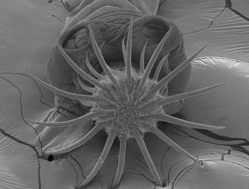
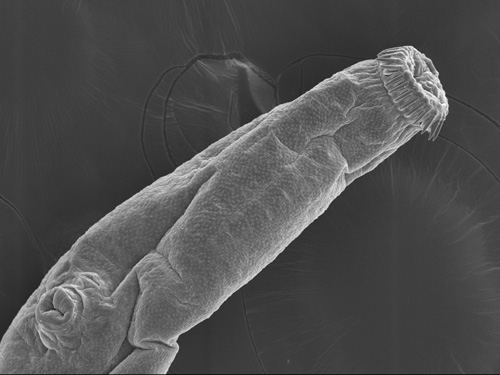
Scanning electron micrograph of the scolex of Paradilepis sp. (Cestoda: Gryporhynchidae). (Photo: Haseeb Randhawa)
See Presswell et al. (2012) Journal of Helminthology 86: 453-464
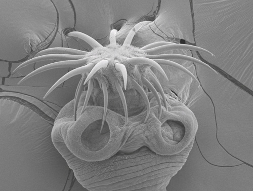
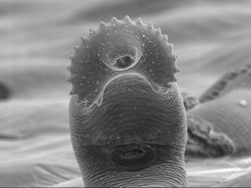
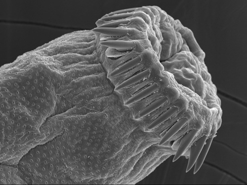
Scanning electron micrograph of the crown of hooks on the rostellum of Paradilepis sp. (Cestoda: Gryporhynchidae), in apical view. (Photo: Haseeb Randhawa)
See Presswell et al. (2012) Journal of Helminthology 86: 453-464
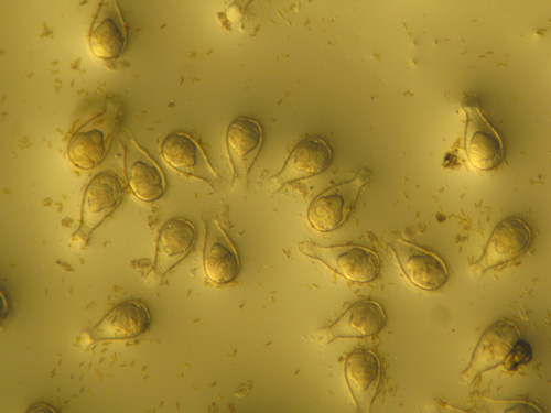
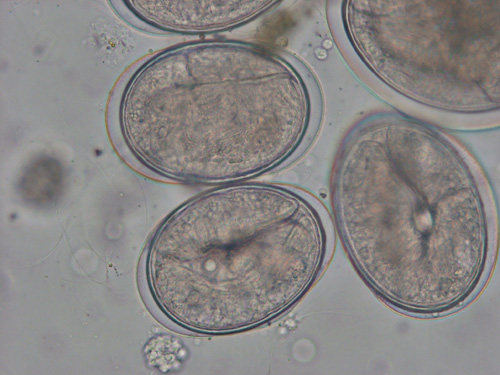
Encysted metacercariae of Philophthalmus sp. (Trematoda: Philophthalmidae), on a microscope slide. (Photo: Fengyang Lei)
See Lei and Poulin (2011) Marine Biology 158: 995-1003
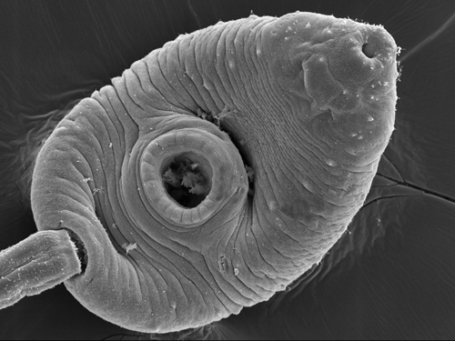
Scanning electron micrograph of the cercarial body of Acanthoparyphium sp. (Trematoda: Echinostomatidae), in ventral view. (Photo: Haseeb Randhawa & Matthew Downes)
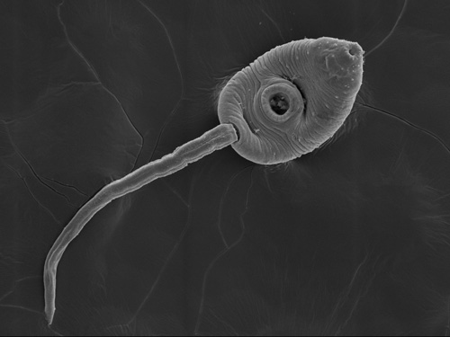
Scanning electron micrograph of a complete cercaria of Acanthoparyphium sp. (Trematoda: Echinostomatidae), in ventral view. (Photo: Haseeb Randhawa & Matthew Downes)
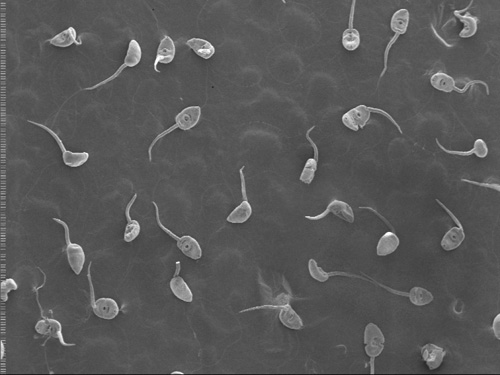
Scanning electron micrograph of numerous cercariae of Acanthoparyphium sp. (Trematoda: Echinostomatidae). (Photo: Haseeb Randhawa & Matthew Downes)
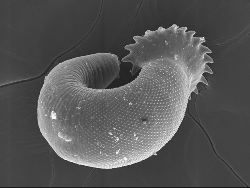
Scanning electron micrograph of an excysted metacercaria of Acanthoparyphium sp. (Trematoda: Echinostomatidae), in dorsal view, taken from its cockle second intermediate host. (Photo: Haseeb Randhawa & Matthew Downes)
Scanning electron micrograph of an excysted metacercaria of Acanthoparyphium sp. (Trematoda: Echinostomatidae), showing the collar spine in ventral view, taken from its cockle second intermediate host. (Photo: Haseeb Randhawa & Matthew Downes)
Scanning electron micrograph of the cercarial body of Curtuteria australis (Trematoda: Echinostomatidae), in ventral view. (Photo: Haseeb Randhawa & Matthew Downes)
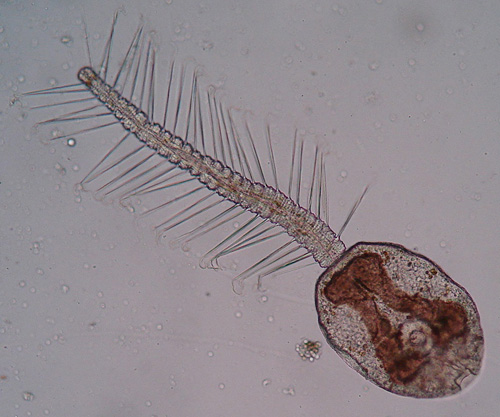
The rarely found Cercaria pectinata (Trematoda), which uses the New Zealand cockle as its first intermediate host. (Photo: Tommy Leung)
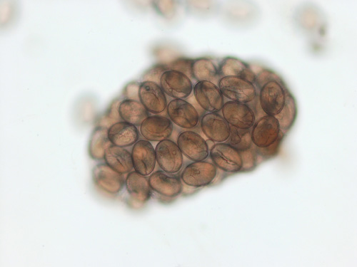
Encysted metacercariae of a microphallid trematode, within a sporocyst, inside the pulmonate snail Amphibola crenata. (Photo: Tommy Leung)
Close-up view of encysted metacercariae of a microphallid trematode, within a sporocyst, inside the pulmonate snail Amphibola crenata. (Photo: Tommy Leung)
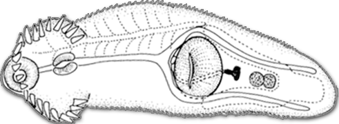
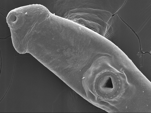
Scanning electron micrograph of an excysted metacercaria of Telogaster opisthorchis (Trematoda: Cryptogonimidae), taken from a fish second intermediate host. (Photo: Haseeb Randhawa & Matthew Downes)
Scanning electron micrograph of the oral sucker, and its ring of spines, on an excysted metacercaria of Telogaster opisthorchis (Trematoda: Cryptogonimidae). (Photo: Haseeb Randhawa & Matthew Downes)
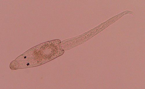
Monostome cercaria emerged from the freshwater snail Potamopyrgus antipodarum. (Photo: Otago Parasitology Group)
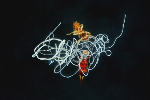
Undescribed mermithid nematodes issued from an earwig caught in the Dunedin Botanical Gardens. (Photo: Haseeb Randhawa & Ken Miller)