Tuesday 17 July 2018 9:13pm
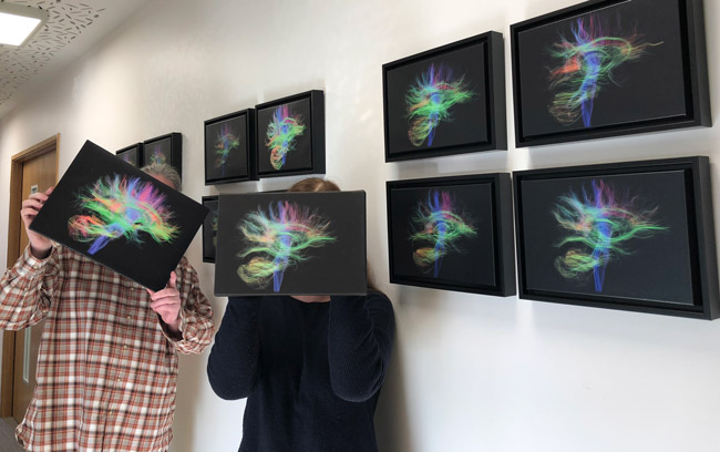
Sean Hogan and Karen Tustin hold the MRI scans of their own brains - which have been printed and displayed on the wall of the Dunedin Study building.
The team at the University’s Dunedin Multidisciplinary Health & Development Research Unit (DMHDRU) is displaying staff photos with a difference – scans of their own brains.
Administration and Communications Officer Jenny McArthur explains that the Dunedin Study includes a new MRI Brain Imaging Study in the 45-year-old cohort testing, and that before that phase began team members had a scan as “guinea pigs”.
Those staff scans have been printed on canvases and now adorn the walls of the building. Mrs McArthur says since they went up they’ve received a great reaction from visitors to the building.
“We’ve also had a competition to see if people could guess whose was whose,” she laughs. “But really it’s impossible to guess.”
The “Dunedin Study” is the most detailed of its kind in the world. It has followed a cohort of 1037 children born at Queen Mary Maternity Hospital in 1972-73 in Dunedin, through their childhoods and into middle age.
The 45-year-old testing began in August 2016, and will finish in December.
The new Brain Imaging Study is gathering information to help scientists understand how parts of the brain influence people’s reaction to different experiences in their daily life.
The results will be linked back to all of the other information gathered by the study to see if people’s experiences in early life might shape their brain function and how this might influence their life as adults.
The scans will also help scientists learn more about how the brain is connected to those parts of the body’s functions which are involved in diseases of lifestyle and age.