Imaging Facilities
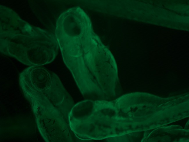
The development of zebrafish with specific tissues or organs of interest marked as fluorescent enhances the study of the zebrafish's embryology, which is already highly visible due to the transparent embryos.
The Otago Zebrafish Facility stocks several lines of fluorescent zebrafish, where different tissues are marked green (GFP) or red (RFP). These can then be studied under the OZF's suite of imaging equipment which currently includes the following:
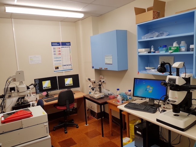
- Nikon C2 Automated Confocal Microscope Nikon's Eclipse Ni-E is fully motorised and can image fluorescence in a number of channels. Through the attached microscope camera kit, researchers can capture 2- and 3D imagery for publication purposes and record movie clips.
- Leica M205 FA Fluorescent Stereomicroscope Fully motorised and is one of the highest resolving power and largest zoom range microscope commercially available. The Leica can view fluorescence in three channels – UV, GFP and dsRed. Through a microscope camera kit, researchers can capture 2- and 3D imagery for publication purposes and record movie clips.
- Leica Stereo M80 Microscopes x 2
- Leica Stereo MZ75 Microscope
- Nikon SMZ1500 Stereoscopic Zoom Microscope with a digital camera kit