
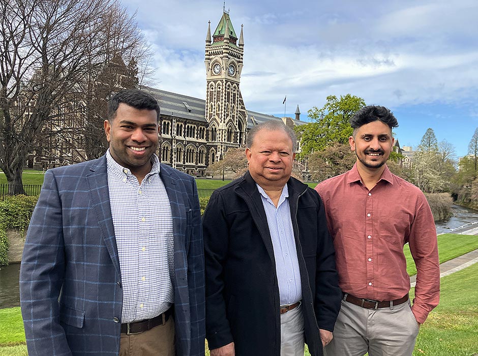
University of Otago – Ōtākou Whakaihu Waka Health Sciences researchers, from left, Dr Jithendra Ratnayake, Professor George Dias and Dr Niranjan Ramesh have developed and patented a new bone graft process.
University of Otago – Ōtākou Whakaihu Waka researchers have developed a new patented process that could improve the way bone graft materials are produced for medical and dental applications.
The technology, developed in a Department of Anatomy laboratory led by Professor George Dias in Dunedin, uses cow bone to produce hydroxyapatite, a naturally occurring mineral that makes up much of the hard structure of human bones and teeth.
The process has now been patented.
Bone grafts are used to repair bone defects resulting from fractures, for tumour removal, and for a range of dental procedures such as wisdom tooth extraction or for dental implants.
George explains that bone grafting is already widely used in medicine and dentistry, but existing approaches have limitations.
One of the traditional options is an autograft, where bone is transferred from another part of the patient’s body. This means an extra surgical procedure, which can increase operating time, cost, and patient discomfort, and risks complications at the bone-harvesting site.
Another graft alternative is synthetic bone substitutes. However, it’s challenging to manufacture materials that closely replicate the complex composition and porous architecture of natural bone. So to work around this, scientists add selected ions to the laboratory-made synthetic hydroxyapatite.
Natural cow bone is a meat processing by-product in New Zealand. It contains trace elements which can contribute to bone healing.
George says the ideal bone tissue engineering solution has always been to integrate the natural advantages extracted from bovine bone with the high bioactivity of synthetic hydroxyapatite.
But that has proved tricky. Because ion substitution is so chemically difficult, no-one had come up with a way to incorporate beneficial ions into bovine bone-derived hydroxyapatite while still maintaining the porous structure.
It was a challenge George and his University of Otago research team, including Dr Jithendra Ratnayake and Dr Niranjan Ramesh – then PhD candidates – took on and won.
Jithendra’s PhD thesis in 2017 focused on developing a bone graft substitute from waste bovine bone and incorporating fluoride and silicate ions into bovine bone-derived hydroxyapatite.
The aim was to develop two bone graft substitutes and investigate their chemical, physical, and biological properties.
He and George spent 18 months of trial and error to find a way to incorporate a single ion into bovine-derived hydroxyapatite.
“Working through different procedures was hard, and there were times we wanted to walk away," Jithendra says.
“But taking the results into George’s office when we’d finally figured it out was the most exciting moment.”
Their process uses a chemical method to introduce selected ions into hydroxyapatite produced from bovine bone.
This is all done without destroying the microscopically tiny architecture that makes natural bone regenerate.
“The approach brings together the advantages of both natural and engineered bone graft materials,” George says.
Then they worked with Niranjan on double ion substitution techniques, another laborious and time-consuming process which also took many months to develop.
Niranjan’s PhD research in 2020 focused on co-substituting ions (strontium and magnesium) into bovine bone-derived hydroxyapatite.
He successfully developed a co-substitution process that incorporated both ions, which is a significant breakthrough.
“The challenge involved in the co-substitution process was improving the functionality of the bovine hydroxyapatite without compromising on its structural properties.
"With time, we were able to successfully build on Jithendra’s work and advance the process,” Niranjan says.
“At the end of the day it’s about the patients – the benefits are considerable for those needing bone grafts. We’re also getting great feedback from doctors, dental specialists and health professionals working with the new product." – Professor George Dias
George, Jithendra and Niranjan have taken their development to the commercial stage and are currently working with a biotechnology company in India that has patented the novel process in the United States.
The Colombo-based company has been testing the base bone hydroxyapatite product and organising regulatory approvals; this base product has already been trialled in bone hydroxyapatite grafts and root canal work in dental surgery.
Now they are using the technique to produce a granule product for other medical applications, and are looking at larger scale commercial manufacturing in Sri Lanka, using bone sourced from New Zealand.
It took over six years, but George says seeing the product in the market benefitting people needing dental and maxillofacial work is hugely rewarding.
“At the end of the day it’s about the patients – the benefits are considerable for those needing bone grafts. We’re also getting great feedback from doctors, dental specialists and health professionals working with the new product.
It’s also a great feeling to see success coming from a small group in a small country,” he says.
“We’re proof that you can work smartly with a small amount of investment, and that we can collaborate successfully with companies that can take it further. And also, it’s great to use bone which is a byproduct in the New Zealand cattle meat industry.”
Jithendra is now a senior lecturer in the Otago Faculty of Dentistry, working on developing biomaterials from sustainable waste products. Niranjan is a Teaching Fellow in the Department of Anatomy, teaching in the Health Sciences First Year programme and 200-level anatomy courses.
Why the scientists are using bovine
Hydroxyapatite is used as a bone substitute. It is biocompatible, meaning it can be well tolerated by the body, and bioactive, meaning it can interact with surrounding tissue and support new bone formation.
Hydroxyapatite derived from bovine bone has had an important advantage over the synthetically made version – after appropriate processing, it retains the naturally porous structure.
George explains these tiny, interconnected spaces provide pathways through which cells, blood vessels and new tissue can grow as the damaged area heals.
The small amounts of trace elements found in natural bone, such as magnesium, strontium and silicate, can contribute to bone healing.
The other advantage of bovine bone is that it is a readily available by-product – rather than allowing it going to waste, it can be converted into a higher-value biomedical product.
George and Jithendra have also looked at other bone sources. Chicken bones are not strong enough, but Jithendra has published a paper showing the process successfully produced from camel bones sourced from Saudi Arabia.
Their work was supported by the University of Otago’s Research and Enterprise office.
– Kōrero by Claire Grant, Communications Advisor, Faculty of Biomedical Sciences
Department of Anatomy
Find out more about the Department of Anatomy's studies into biomaterials, clinical anatomy and forensic science.
Anatomy research