Below is a description of the major items of equipment available. We strongly encourage you to seek our advice in the early planning of your project.
To use our equipment you will need to register your project and undergo training.
Confocal microscopy registration
For advice contact:
Email confocal.microscopy@otago.ac.nz
Zeiss LSM+ 900 with Airyscan 2
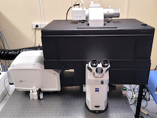
The Zeiss LSM 900 is a compact confocal microscope for high resolution, live cell imaging. The system offers 3 channel simultaneous imaging, enhanced resolution Airy scanning and spectral imaging. Post-acquisition processing with Airy scanning, Joint Deconvolution or LSM Plus allows users to achieve up to 90nm resolution without compromising their sample. Using the 900+ multiplex modes imaging speed can be increased to 18 fps (full frame) for dynamic live cell studies.
This microscope is equipped with an incubation chamber allowing users to environmentally control the conditions for long duration live cell imaging. The Zeiss Zen software is very intuitive with the smart setup function making this an easy microscope to use.
Applications
- Super resolution imaging
- Full environmental controls
- Time-lapse imaging
- Multipoint time series
- Montaging/large images
- FRAP/FLIP
- FRET
- Spectral unmixing
- 3D imaging with jDCV
Specifications
Illumination
- Colibri 5 type RGB-UV 4-channel fluorescence light source
Detectors
- 2 spectral detector channels, GaAsP PMT with 8 bit and 16 bit dynamic range
- 1 Airyscan detector for 63x objective
Objectives
- 10x Air (0.45 NA)
- 20x Air (0.8 NA)
- 40x Water (1.2 NA)
- 63x Oil (1.4 NA)
Laser (solid state) Excitation Lines
- 405nm
- 488nm
- 561nm
- 640nm
Emission Filters
Variable secondary beam splitter (VSD) with short pass (SP) and long pass (LP) filters
Maximum Frame Resolution
- 6144 x 6144 pixels
- 4096 x 4096 pixels with Airyscan
Software
Zen 3.7
Charges
$65 per hour
Tier Fees
Bookings
Zeiss LSM+ 900 with Airyscan 2 booking
Airyscan Information Link
Andor Dragonfly – Spinning Disk Microscope
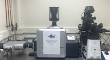
The Andor Dragonfly is a dual microlens spinning disk confocal microscope. It captures images 10x faster than a conventional confocal microscope, with improved sensitivity. This three camera confocal microscope includes widefield, confocal, TIRF, laser ablation, 3D STORM and 2-colour simultaneous confocal imaging modalities.
The Dragonfly is ideal for live cell imaging providing low phototoxicity and photobleaching. It has a large field of view which makes it perfect for imaging live cell or fixed samples. Borealis illumination provides stability, throughput, uniformity and extended wavelength range.
Applications
- Widefield or confocal imaging modes
- Simultaneous multi-colour TIRF
- SRRF-Stream
- dStorm
- Micropoint
- Time-lapse imaging
- Multipoint time series
- Montaging/large images
- 3D imaging
- Environmentals available
Specifications
Cameras
- iXon EMCCD, 1024x1024 pixels, 13µm pixel size
- 2 x Zyla 4.2 sCMOS, 2048x2048 pixels, 6.5µm pixel size
Objectives
- 10x Air (0.45 NA)
- 20x Air (0.75 NA)
- 40x Air (0.95 NA)
- 60x Water (1.2 NA)
- 60x Oil (1.49 NA) TIRF
- 100x Oil (1.45 NA)
Confocal Pinhole Diameter
- 25µm
- 40µm
Laser (solid state) Excitation Lines
- 405nm
- 488nm
- 561nm
- 640nm
Emission Filters
- 450±50nm
- 521±38nm
- 600±50nm or 620±60nm
- 700±75nm or 698±70nm
(specialised filter set available for Zyla Camera)
Software
Fusion with real-time 3D rendering
Charges
$65 per hour
Tier Fees
Bookings
Andor Dragonfly – Spinning Disk Microscope booking
Opera Phenix High-Content Screening System
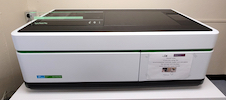
The Opera Phenix is a microlens-enhanced spinning disk confocal high content microscope. Including both confocal and widefield fluorescence capabilities the Opera Phenix can automatically scan multi-well plates and slides under a range of magnifications from 4x to 63x in 3D and as a time series.
Complex and data rich images can be processed through a free browser driven app (Signal Image Artist) to measure thousands of parameters per image/well/plate using Ai trained segmentation routines for a high content analysis of entire experiments in a single operation. The Opera Phenix is equipped with environmental controls for live cell experiments this includes: temperature control (37-42ºC ± 1ºC), CO2 control and humidity control.
Applications
- Widefield or confocal imaging modes
- Digital phase contrast imaging
- Preciscan
- High-throughput high-content assays
- Environmentally controlled for live cell experiments
- Phenotypic screening
- Time-lapse imaging
- Multipoint time series
- Montaging/large images
- 3D imaging
- Ratiometric FRET
Specifications
Camera
sCMOS sensor up to 100fps, with 16 bit resolution.
Objectives
- 5x Air (0.16 NA)
- 10x Air (0.3 NA)
- 20x Air (0.4 NA)
- 63x Water immersion (1.15 NA)
Laser (solid state) Excitation Lines
- 405nm
- 488nm
- 561nm
- 640nm
Emission Bandpass Filters
- 435-480nm
- 500-550nm
- 570-630nm
- 650-760nm
Resolution
2160 x 2160 pixels, 6.5μm pixel size
Software
- Harmony 5.1 (With Phenologic)
- Signals Image Artist (online data analysis)
Charges
$65 per hour
Tier Fees
Bookings
Opera Phenix High-Content Screening System booking
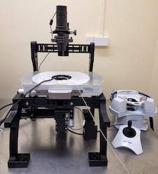
Neoscan N80 Micro-Computed Tomograph (µCT) microscope
NEOSCAN N80 is a desktop, general purpose X-ray laboratory instrument for non-destructive 3-dimensional reconstruction of internal microstructure of the objects with spatial resolution in the micron range. Typical applications include, but are not limited to, a medium-large size plastic molded or 3D printed parts, light metals (Al, Ti) and small parts in more dense metals (steel, …), ceramics, carbon- and glass-fiber composites, biological materials (tissue samples, bones, implants, …), wood, electronics, glass, geological samples – plugs and drilled cores, pharmaceutical products and packaging, etc.
Applications
- 3D X-Ray imaging for a wide range of materials from dense metal to soft tissue
- 8 preinstalled and calibration filters to suit most material
- Ring Artefact free fat panel detector
- Quantitate bone density measurement (measured against sets of BMD phantoms)
- Helical scanning
- Licence free acquisition and analysis software
Specifications
General
- Pixel size at maximum magnification - <1.2 μm
- True low-contrast 3D resolution - 2 μm or better
- Maximum scanning diameter - 100 mm
- Maximum scanning length - 163 mm
- Maximum physical object length - 200 mm
X-Ray Source
- Maximum voltage - 110kV
- Maximum power - 16W
- Smallest spot size - 2 μm or smaller
- Automatic filter changer, number of positions – 20
X-Ray Detector
- Image sensor - 7 Mp Flat Panel
- Scintillator - GADOX
Integrated options
Micro-positioning stage (travel) - Integrated (10 mm)
Software
- Neoscan N60
- Dragonfly
Charges
$65 per hour
Tier Fees
Bookings
NeoScan N80 Links
https://neoscan.com/system/neoscan-n80/
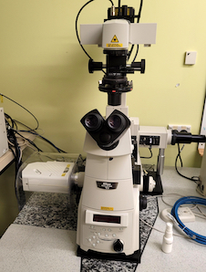
Nikon A1+ Inverted Confocal Laser Scanning Microscope
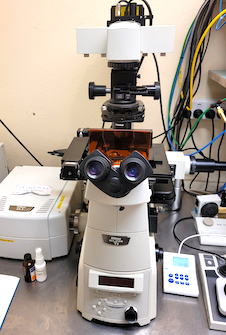
The Nikon A1+ is a point scanning confocal on an inverted microscope base which can achieve both high-quality images for spatial information and high-speed images of fast-moving events. It is equipped with a galvano scanner enabling high resolution imaging of up to 4096 x 4096 pixels. With the addition of high-sensitivity gallium arsenide phosphide (GaAsP) detectors the A1+ can achieve higher quantum efficiency resulting in brighter imaging with minimal noise.
This microscope has an automatic focus mechanism – Perfect Focus System (PFS) which continuously corrects focus drift during long time-lapse observation. The A1+ is equipped with deconvolution in both 2D and 3D within NIS Elements Software.
Applications
- Confocal microscopy
- Differential interference contrast
- Time-lapse imaging
- Multipoint time series
- Montaging/large images
- Z-Series
- FRAP
- FRET
Specifications
Illumination for Fluorescence
pE300 light source
Objectives
- 10x Plan (0.45NA)
- 20x Plan Apo (0.8 NA)
- 40x Plan Fluor oil immersion (1.3 NA)
- 60x Plan Apo oil immersion (1.42 NA)
Laser (solid state) Excitation Lines
- 405nm
- 488nm
- 561nm
- 640nm
Filter Sets
- Emission 450 ± 50nm
- Emission 525 ± 50nm
- Emission 595 ± 50nm
- Emission 700 ± 75nm
Detectors
- 2 PMT Detectors
- 2 GaASP Detectors
Resolution
4096 x 4096 pixels
Software
NIS Elements High Content Package (version 5.02) with deconvolution
Charges
$65 per hour
Tier Fees
Bookings
Nikon A1+ Inverted Confocal Laser Scanning Microscope booking
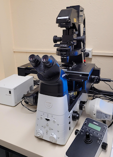
Bruker BioScope Resolve AFM
The Bruker Bioscope Resolve is an Atomic Force Microscope mounted on an inverted microscope platform. This enables correlation between both atomic force and optical (brightfield and fluorescence) imaging. The AFM can operate under traditional contact and tapping AFM modes together with Peak Force Tapping.
With an imaging resolution approaching 5nm (in height) the Bioscope resolve can operate in both air and liquid environments to capture both topographical and surface mechanical features of a range of materials. The Peak Force Tapping mode forms the core of ScanAsyst to automate parameter optimization to greatly improve ease of use, and PFQMN for quantitative measurement of mechanical properties of your samples , including living cells.
Applications
AFM
- Topographical AFM
- Fluid imaging AFM
- Live cell AFM
- Correlative AFM/Fluorescence
- Quantitative nanomechanical mapping
- Fast force volumes
Specifications
AFM mode
- Contact mode AFM
- Tapping Mode AFM
- PeakForce Tapping AFM
- Nanomechanical mapping (PFQNM)
- Fast force volume and single force curves
- MIRO (Microscope Image Registration and Overlay) AFM imaging
Optical
Illumination
- Diacopic illumination vis 535nm LED
- Fluorescence via CoolLED pE300
Detectors
Pco. Panda 26 sCMOS camera
Objectives
10x -60x depending on requirement (using single objective nosepiece)
Emission Filters
- Pos 1: Ex 350/80nm ; Em 420/40nm
- Pos 2: Ex 470/40nm ; Em 515/20nm
- Pos 3: Ex 557/55nm ; Em 615/50nm
- Pos 4: Brightfield
Software
- Nanscope V - AFM
- Micromanager (for optical only)
Charges
$65 per hour
Tier Fees
Bookings
Bruker BioScope Resolve AFM booking
BioScope Resolve Information Link
AFM
Probes
https://www.brukerafmprobes.com/c-15-probes.aspx
Nikon C2+ Confocal Microscope
The Nikon C2+ is basic confocal microscope however still capable of achieving diffraction limited resolution. Using the motorized Nikon Ti-E, high resolution multichannel 3D images can be automatically captured from a wide range of sample formats. The high-speed galvano scanners, operating at rates of up to 5 fps, enable even the fast-beating motion of cardiac muscles to be captured with precision. The system also provides simultaneous acquisition of three fluorescent channels plus DIC in a single scan.
Applications
- Confocal microscopy
- Differential interference contrast
- Time-lapse imaging
- Multipoint time series
- Montaging/large images
- Z-Series
- FRAP
- FRET
Specifications
Illumination
Epi-Fluorescence using CoolLED pE300
Detectors
Insert
Objectives
- 10x Plan Apochromat Lambda (NA 0.45, WD 4mm)
- 20x Plan Apochromat (NA 0.8, WD 0.8)
- 40x Plan Apochromat Lambda (NA 0.95, WD 0.21)
- 60x Plan Apochromat Lambda S (0.95, WD 0.21)
Laser (solid state) Excitation Lines
- 405nm
- 488nm
- 561nm
Emission Filters
- Dual DAPI/Cy5
- Combined 525/50, 595/40
Maximum Frame Resolution
- Max pixels: 2048x2048
- Max speed: 2 fps (512x512)
Software
NIS Elements
Charges
$30 per hour
Tier Fees
Bookings
Nikon C2+ Confocal Microscope booking
Nikon C2+ Confocal Information Link
https://www.microscope.healthcare.nikon.com/products/confocal-microscopes/c2
Nikon Ti2E Inverted Fluorescence Microscope
The Eclipse Ti2 is an inverted fluorescence microscope offering a 25mm field of view which maximises the sensor area of the large-format CMOS cameras, improving data throughput.
This fully motorised microscope can automatically capture multi-position, multi-channel images using both bright-field (transmitted and DIC) illumination) and multi-channel fluorescence imaging using a range of customised software interfaces. With the addition of both Z-drive and PFS autofocusing systems, to improve system stability, the Ti2-E is a flexible and fast fluorescence microscope for a broad range of applications.
Applications
- Brightfield microscopy
- Differential interference contrast
- Phase contrast
- Fluorescence microscopy
- Time-lapse tissue imaging
- Multipoint time series
- Montaging
- Z-series
Specifications
Illumination
pE300 light source
Objectives
- 10x Plan Apo (0.45NA)
- 20x Plan Apo (0.8 NA)
- 40x Plan Apo (0.95 NA)
- 60x Plan Apo water immersion (1.2 NA)
Filter Sets
- Excitation 395nm : Emission 435 ± 50nm
- Excitation 460-500nm : Emission 510 ± 50nm
- Excitation 527-553nm : Emission 577–633nm
- Excitation 620-650nm : Emission 700–770
Cameras
- Nikon Digital Sight DS-Qi2 CMOS sensor monochrome 7.3MP camera
- Nikon Digital Sight DS-Ri2 CMOS sensor colour 16MP camera
Software
NIS Elements AR (Advanced Research) featuring JOBS module
Charges
$65 per hour
Tier fees
Bookings
Nikon Ti2E Inverted Fluorescence Microscope bookings
Confocal Unit Analysis PCs
Software available
- NIS Elements AR 5.21.03
- Arivis Vision 4D
- Zeiss Zen 3.8
- Harmony 5.1
- FIJI (imageJ)
- Dragonfly
- Neoscan60
- ilastik
- CTAn
- Nrecon
- Dataviewer
- Meshlab
- Slicer 5.0.3
- Volocity