
The Light and Confocal Microscopy Unit is the core imaging facility for light microscopy at the University of Otago.
Based in the Lindo Ferguson Building, we provide access, training and support for an advanced suite of instruments covering light and confocal microscopy, high content screening, super-resolution microscopy, x-ray tomography (microCT) and atomic force microscopy together with advanced imaging analysis.
Your project
Getting started with your project
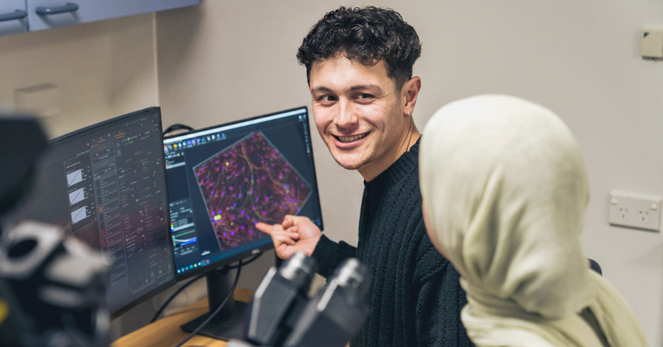
If you’re new to microscopy or are unsure which microscope you need, please arrange a visit with the microscopy team. They will be happy to discuss your project, provide advice on sample preparation and arrange your initial training session.
Contact the team
Dr Rob Woolley
Facility Manager
Email confocal.microscopy@otago.ac.nz
Email rob.woolley@otago.ac.nz
Tel +64 3 479 7308
Rose Smither
Imaging Facility Technician
Email roseanna.smither@otago.ac.nz
Project registration
Project discussion with staff is essential prior to registration and training.
Staff will help you design a practical experiment and provide full training on the equipment. You are not expected to master every function of the instruments and can ask for help at any time, whether it be in using the equipment, the design of your experiment or with image analysis.
Before starting, please read and agree to our user policy and complete a project registration form.
- Confocal Microscopy User Policy (PDF)
- Out of hours user guidelines (PDF)
- OMNI confocal project registration form
Initial advice and consultation
Contact the manager, Dr Rob Woolley, for initial advice and consultation:
Email confocal.microscopy@otago.ac.nz
For advice on the latest academic advances contact:
Dr Laura Gumy
Academic Leader, Confocal Microscopy
Email laura.gumy@otago.ac.nz
Services and techniques
- Access for downloading your confocal data
- Confocal microscopy equipment for trained independent users
- Confocal microscopy image analysis
- Research design advisory services and support
- Training for equipment use
Equipment
The unit hosts a range of imaging systems; therefore we strongly encourage you to seek our advice in the early planning of your project.
Please contact Rob (Woolley) or Rose (Smither) for advice on system capabilities and costs:
Email confocal.microscopy@otago.ac.nz
To use our equipment you will need to register your project and undergo training.
See our confocal microscopy equipment and specifications page or select the relevant links below for all the equipment listed here, with specifications.
Light microscopy
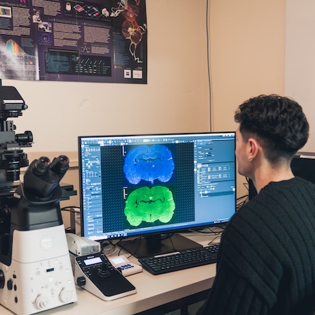 The Nikon Ti2-E inverted microscope offers brightfield, DIC, polarisation and fluorescence imaging modalities with dual cameras for sensitive monochrome and rapid colour imaging. It’s compatible with most sample formats (slides, petri dishes etc), and is fully motorised microscope for large-scale image montages or automated image acquisition.
Nikon Ti2E specifications can be found on our equipment page
Confocal microscopy
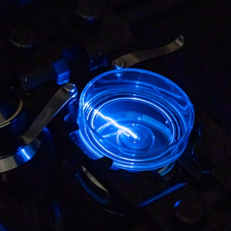 For more advanced imaging applications, the unit hosts 5 confocal microscopes, each offering specific performance advantages and modalities.
Andor Dragonfly Spinning Disk Confocal Microscope
- High-sensitivity confocal: EM-CCD (iXon888)
- Fast confocal imaging: sCMOS
- Simultaneous 2-channel confocal (includes TIRF)
- Simultaneous laser ablation and confocal imaging
- Super-resolution radial fluctuation microscopy (SRRF-Stream)
- Total Internal reflection microscopy (TIRF)
- 3D Single Molecular localisation microscopy (SMLM)
- Live cell imaging
Zeiss LSM 900 Airy2 Confocal Microscope
- Confocal and Airy scanning high resolution
- Spectral Imaging
- FRET
- Live cell imaging
Perkin Elmer Opera Phenix
- High-content widefield and confocal imaging for multi-well plates
- Live cell imaging (PC2)
- Online analysis software (Signals Image Artist)
Nikon A1+ Confocal Microscope
- Advanced resolution (Nikon C-ER deconvolution)
- Simultaneous 4 channel confocal imaging
- FRET
Nikon C2+ Confocal Microscope
- Low-cost confocal imaging
- Simultaneous 3 channel confocal
- FRET
Visit our equipment and specifications page
MicroCT X-Ray scanning
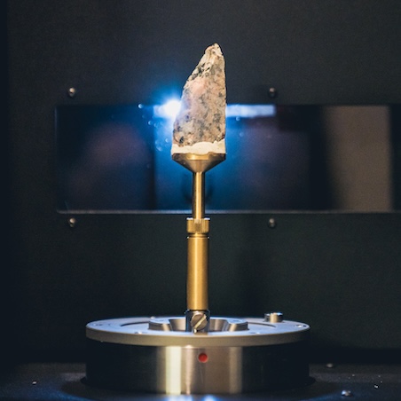 Micro-computed tomography (Micro-CT) uses X-Ray illumination to generate detailed 3-dimensional scans of the internal structures of dense material such as bone, teeth, wood or rocks. The internal composition and density of material can be directly measured and visualised down to 2µm.
- Large sample chamber: 100mm x 220mm (scanning 100m x 130mm object)
- Fast flat-panel CMOS detector
- 110kV 16W Hamamatsu X-Ray source
- 6 emission filters
- Round, multi-position and helical scanning
- 2µm pixels spot (high resolution mode)
Atomic Force microscopy
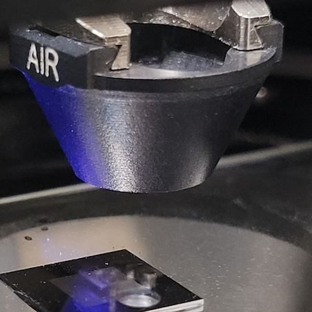 Multiple imaging modalities (Contact, Tapping, Peak Force tapping, PFQMN, force mapping)
- Liquid imaging compatible
- Quantitative nanomechanical mapping (PFQNM)
- Live cell compatible
- Simultaneous AFM+ brightfield and fluorescence imaging
- Correlative AFM + optical imaging (MIRO)
Digital Analysis Suite
- 4 advanced analysis desktop computers
- Comprehensive collection of freeware and commercial image analysis programs
- Latest GPU’s for machine and deep-leaning training routines
- Compatible for processing large and complex datasets
Project registration
Before you can be trained to use any of our equipment, or begin a new project, you must read and agree to our User Policy and complete a Project Registration form.
User Group
The User Group consists of members that represent University of Otago departments that use the Confocal Microscopy unit and it advises the OMNI Academic Advisory Group on direction and strategy.
Visit the About us page for information about how our user groups work and how to make contact.
Costs
Users can choose to be charged at the casual hourly rate or to prepay for a Tiered Fee.
Health Science Division (HSD) rates only apply to HSD Dunedin campus departments.
Tiered fee
Tier user application form (PDF)
OMNI Electron and Confocal Microscopy tiered charges 2025 (PDF)
Training and online resources
Technique training and support
Contact us for training:
Email confocal.microscopy@otago.ac.nz
Image analysis resources and support
Visit confocal microscopy image analysis for advice on programs' capabilities and links to online resources.